GO:0061298Ontology (GO BP)GO biological process · ~19 member genes
Q-omics provides the Retina vasculature development in camera-type eye (GO:0061298) pathway profile, scoring each patient from the combined activity of its roughly 19 member genes. Pathway activity is associated with patient survival in 26 of 34 cancer types, with the highest sampling consensus in LUAD. Among the 18 cancer types available for tumor–normal comparison, the pathway is differentially active in 11, with the highest sampling consensus in BLCA. Additionally, pathway RNA activity shows 34,688 significant cross-omics associations, again with the highest sampling consensus in STAD. Together, these results highlight LUAD, BLCA, and STAD as cancer lineages where the pathway shows reproducible signals across outcome, tissue activity, and molecular association analyses.
Every result is evaluated using two consensus scores. Sampling consensus measures how consistently a finding is reproduced within a cancer lineage across different conditions. Lineage consensus measures how broadly the result is shared across cancer types, distinguishing pan-cancer signals from lineage-specific patterns. Pathway-against-pathway and pathway-against-mutation comparisons are not available for ontology entities.
Survival associations
This table summarizes Retina vasculature development in camera-type eye survival associations by molecular data type. RNA-level pathway activity shows survival associations in the most cancer types (26). The rightmost column indicates the cancer type with the highest sampling consensus for each layer.
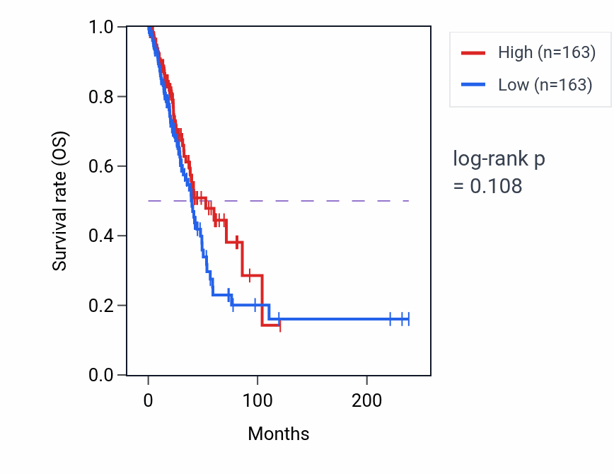
This table ranks reproducible pathway activity–survival associations across cancer types. High Retina vasculature development in camera-type eye activity shows favorable associations in LUAD, HNSC and THCA, but unfavorable associations in KIRP, UVM and MESO. In the LUAD Kaplan–Meier curve the low-activity group declines faster, consistent with the favorable association (log-rank p < 0.001). LUAD ranks highest by sampling consensus for Retina vasculature development in camera-type eye.
This table summarizes Retina vasculature development in camera-type eye tumor–normal activity differences by data type. RNA-level activity shows significant tumor–normal differences in 11 cancer types, while mass-spec protein activity shows differences in 2. The strongest signals are in BLCA for RNA and LUAD for protein.
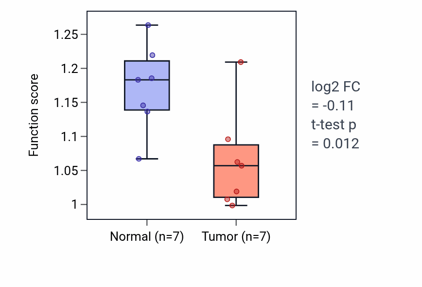
This table ranks reproducible tumor–normal activity differences for the pathway. A positive fold-change indicates higher activity in tumor tissue. The pathway shows consistently lower tumor activity across BLCA, KICH, LUAD, KIRP, LUSC and UCEC. In the BLCA box plot, normal samples show higher pathway activity than tumor samples (log2 FC = −0.125, t-test p < 0.001).
This table shows molecular features associated with Retina vasculature development in camera-type eye pathway activity in patient tissues and cancer cell lines. In patient samples, pathway activity is most strongly linked to RNA and protein features, with the largest associated set in STAD. In cancer cell lines, RNA-expression features and functional dependencies dominate, with the largest set in OESOPHAGUS.