MIR1-1HG
RNA — tumor vs normalAcross TCGA pan-cancer cohorts, MIR1-1HG RNA differs between tumor and matched normal tissue in 8 of 18 cancer types tested, making tumor–normal expression one of MIR1-1HG’s most consistent transcriptional readouts.
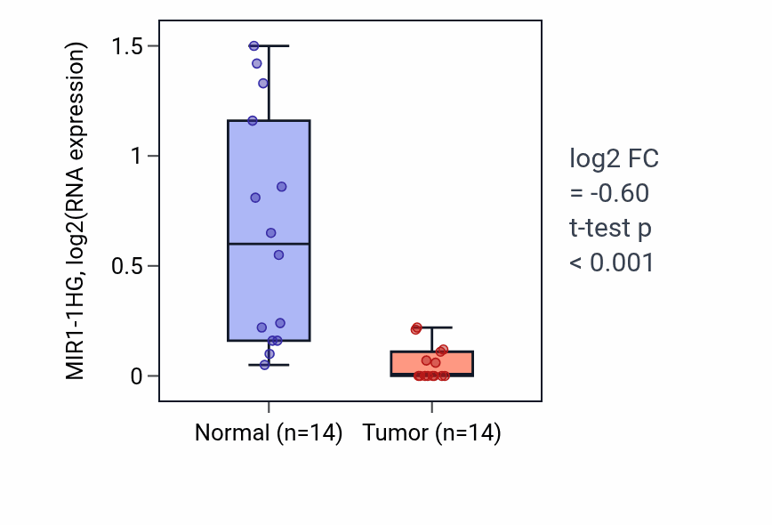
The strongest signal is observed in bladder urothelial carcinoma (BLCA), where MIR1-1HG RNA is repressed in tumor relative to normal tissue. In most cancer types MIR1-1HG is over-expressed in tumor, although a few such as BLCA and COAD show the opposite, repressed pattern.
BLCA, COAD, and HNSC are the cancer types where MIR1-1HG tumor–normal differential expression is most reproducible.
RNA tumor vs normal associations by lineage
Ranked by sampling consensus. Fold-change is the tumor-versus-normal difference in MIR1-1HG RNA (log2); positive values indicate higher expression in tumor. p-values are from the differential-expression test.
Pink = over-expressed in tumor, green = repressed in tumor. Showing the 8 strongest of 8 lineages.
Exploration
- Same gene, other analysis: MIR1-1HG RNA expression & survival · all MIR1-1HG data types