PPAT
RNA & survivalAcross TCGA pan-cancer cohorts, PPAT RNA is linked to patient survival in 25 of 34 cancer types, making it the most broadly survival-associated PPAT data layer compared with 4 for mutation status and 5 for mass-spec protein.
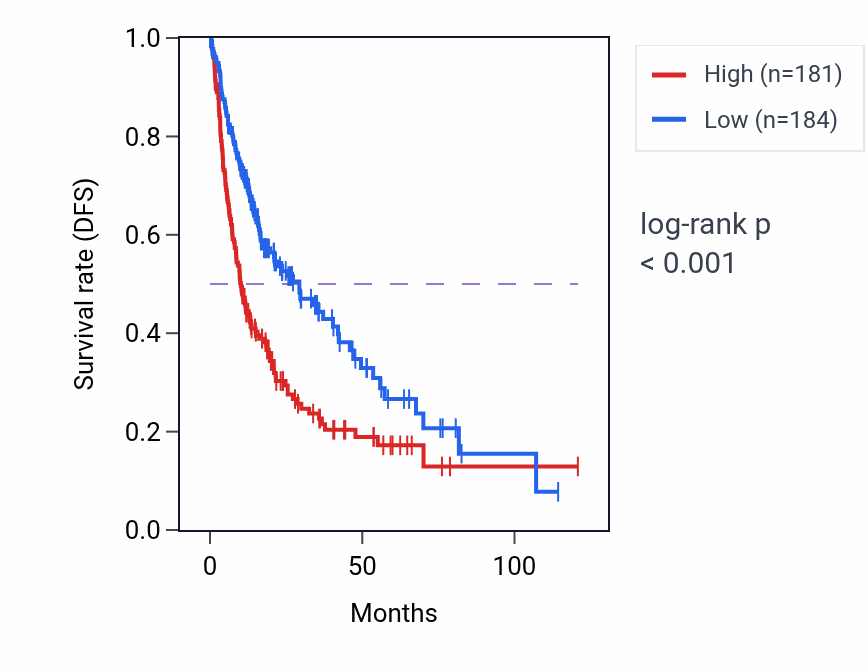
The strongest signal is observed in liver hepatocellular carcinoma (LIHC), where higher PPAT RNA is associated with worse disease-free survival. In most high-consensus cancer types, elevated PPAT expression acts as an unfavorable survival marker, although some lineages such as READ and KIRC show a favorable association.
LIHC, MESO, and READ are the cancer types where PPAT RNA most reproducibly stratifies survival.
RNA survival associations by lineage
Ranked by sampling consensus. AUC1 and AUC2 indicate survival in the high- and low-expression groups, respectively; the lower AUC marks the poorer-surviving group. p-values are from the log-rank test.
Pink = unfavorable, green = favorable. Showing the 12 strongest of 25 lineages.
Exploration
- Same gene, other analysis: PPAT tumor-vs-normal RNA expression · all PPAT data types